Leta i den här bloggen


torsdag 9 oktober 2014

Wnt signalointitiet ja Wnt-proteiinit

 https://www.ncbi.nlm.nih.gov/pubmed/28619731/
 Aberrant activation of the Wnt signaling pathway plays an important role in human cancer development. Wnt signaling is negatively regulated by Axin, a scaffolding protein that controls a rate-limiting step in the destruction of β-catenin, the central activator of the Wnt pathway. In Wnt-stimulated cells, Axin is rapidly modified by tankyrase-mediated poly(ADP-ribosyl)ation, which promotes the proteolysis of Axin and consequent stabilization of β-catenin. Thus, regulation of the levels and activity of tankyrases is mechanistically important in controlling Wnt signaling. Here, we identify ubiquitin-specific protease 25 (USP25) as a positive regulator of Wnt/β-catenin signaling. We found that USP25 directly interacted with tankyrases to promote their deubiquitination and stabilization. We demonstrated that USP25 deficiency could promote the degradation of tankyrases and consequent stabilization of Axin to antagonize Wnt signaling. We further characterized the interaction between TNKS1 and USP25 by X-ray crystal structure determination. Our results provide important new insights into the molecular mechanism that regulates the turnover of tankyrases and the possibility of targeting the stability of tankyrases by antagonizing their interaction with USP25 to modulate the Wnt/β-catenin pathway.


 23.10.2019 : Wnt-beta-cateniinitien  säätely ubikitiini-proteosomi-järjestelmän avulla

Angers S, Thorpe CJ, Biechele TL, Goldenberg SJ, Zheng N, MacCoss MJ, Moon RT.
Nat Cell Biol. 2006 Apr;8(4):348-57. Epub 2006 Mar 19.
PMID:
16547521


2014

https://www.youtube.com/watch?v=YuVkRTOYOlQ

Wnt-signaalitiet ovat joukko signaalinjohtumisteitä proteiineilla, jotka johtavat  solun ulkopuolelta signaalin soluseinän läpi pintareseptorin avulla solun sisälle. On olemassa kolme Wnt- signaalitietä:
(1)  kanoninen Wnt-tie ( johtaa geenitranskription säätelyyn)
(2) nonkanoninen (planar cell polarity) ( säätää  sytoskeletonia ja vastaa solumuodosta)
(3)  nonkanoninen (Wnt/Ca-tie)(  säätelee solunsisäistä kalsiumia)
Kaikki kolme  aktivoituvat, kun Wnt-proteiini ligandi sitoutuu Frizzled-perheen reseptoriin, joka välittää signaalin Dishevelled proteiinille solun sisään.

The Wnt signaling pathways are a group of signal transduction pathways made of proteins that pass signals from outside of a cell through cell surface receptors to the inside of the cell. Three Wnt signaling pathways have been characterized: the canonical Wnt pathway, the noncanonical planar cell polarity pathway, and the noncanonical Wnt/calcium pathway. All three Wnt signaling pathways are activated by the binding of a Wnt-protein ligand to a Frizzled family receptor, which passes the biological signal to the protein Dishevelled inside the cell. The canonical Wnt pathway leads to regulation of gene transcription, the noncanonical planar cell polarity pathway regulates the cytoskeleton that is responsible for the shape of the cell, and the noncanonical Wnt/calcium pathway regulates calcium inside the cell. 

 Wnt-signaalitiet käyttävät läh solu-solu-kommunikaatiota ( parakriinista) tai samansolun kommunikaatiota ( autokriinista). Ne ovat  mitä suurimmassa määrin evolutionaalisesti konservoituneita  järjestelmiä, mikä tarkoittaa, että ne ovat samanlaisia eri lajeilla hedelmäkärpäsestä ihmiseen.

Wnt signaling pathways use either nearby cell-cell communication (paracrine) or same-cell communication (autocrine). They are highly evolutionarily conserved, which means they are similar across many species from fruit flies to humans.[1][2]

 Wnt-signalointi keksittiin ensi kerran  sen osuudesta syövän syntymiseen, mutta  sittemmin  se on tunnistettu myös  alkioajan kehityksestä.  Alkioajan prosesseissa se kontrolloi  kehoakselimallin muodostumista, solun erikoistumiskohtaloita, solun proliferoitumista ja solujen migroitumista. Tällaiset prosessit ovat tärkeitä kudosten asianmukaiselle muodostumiselle, kuten esim luuston, sydämen ja lihasten muodostumisille.  Wnt-tien  rooli alkioajan kehityksessä  keksittiin, kun Wnt-tien proteiinien mutaatioista  tuottui  epänormaaleja  hedelmäkärpäsalkioita.  Myöhemmin tutkijat  havaitsivat, että    epänormaaliuksen taustalla olleet geenit     vaikuttivat myös rintasyövän kehittymistä hiirellä. Wnt- järjestelmän kliininen tärkeys selveni mutaatiotutkimuksilla, joissa  aiheutui useita  tauteja, mm.  rintasyöpää, prostatasyöpää, glioblastomaa, Ii tyypin diabetesta ja muita tauteja.

Wnt signaling was first identified for its role in carcinogenesis, but has since been recognized for its function in embryonic development. The embryonic processes it controls include body axis patterning, cell fate specification, cell proliferation, and cell migration. These processes are necessary for proper formation of important tissues including bone, heart, and muscle. Its role in embryonic development was discovered when genetic mutations in proteins in the Wnt pathway produced abnormal fruit fly embryos. Later research found that the genes responsible for these abnormalities also influenced breast cancer development in mice.
The clinical importance of this pathway has been demonstrated by mutations that lead to a variety of diseases, including breast and prostate cancer, glioblastoma, type II diabetes, and others.[3][4]

 Wnt- proteiinit ovat  sekalainen perhe  erittyneitä lipidillä modifioituneita signaloivia glykoproteiineja, joissa on 350- 400 aminohapon ketju. Näissä proteiineissa oleva lipidimodifikaatio on tyypiltääm  cysteiinien palmitylaatioita. konservoituneesti   23-24 cysteinyylissä. Palmitylaatio on tärkeä seikka, koska  se alkaa Wnt-proteiinin kohdentamisen plasmakalvoon erittymistä varten ja sallii  Wnt- proteiinin sitoutua reseptoriinsa rasvahappojen kovalentilla liitoksella. Wnt-proteiinit käyvät läpi myös glykosylation, jossa liittyy hiilihydraattia  varmistamaan asianmukaisen erittymisen.  Wnt- signaloinnissa tämä proteiinit toimivat ligandeina aktivoiden erilaisia Wnt- teitä para-tai  autokriinisesti. Kuten  edellä mainittu, nämä proteiinitkin  voat hyvin konservoituneita kauta eri lajien. niitä siis on hiirissä, ihmisissä, Xenobus, Zebrafish, Drosophila ym lajeissa.

The Wnt proteins are a diverse family of secreted lipid-modified signaling glycoproteins that are 350–400 amino acids in length.[7] The type of lipid modification that occurs on these proteins is palmitoylation of cysteines in a conserved pattern of 23–24 cysteine residues.[3] Palmitoylation is necessary because it initiates targeting of the Wnt protein to the plasma membrane for secretion and it allows the Wnt protein to bind its receptor due to the covalent attachment of fatty acids. Wnt proteins also undergo glycosylation, which attaches a carbohydrate in order to insure proper secretion.[8] In Wnt signaling, these proteins act as ligands to activate the different Wnt pathways via paracrine and autocrine routes.[1][4]
These proteins are also highly conserved across species.[2] They can be found in mice, humans, Xenopus, Zebrafish, Drosophila, and many others.[9]
WIKI-Taulukosta on tähän otettu sitaatti vain ihmisen Wnt- proteiineista. 
Species
Wnt proteins
Wingless-type MMTV integration site family members
Homo sapiens Wnt1, Wnt2, Wnt2B, Wnt3, Wnt3A, Wnt4, Wnt5A, Wnt5B, Wnt6, Wnt7A, Wnt7B, Wnt8A, Wnt8B, Wnt9A, Wnt9B, Wnt10A, Wnt10B, Wnt11, Wnt16
Perustavaa  tietoa Wnt- signaloinnista
Wnt-signalointi alkaa, kun yksi Wnt- proteiini sitoutuu Fz- perheen reseptorin  N- terminaaliseen ja   solun ulkopuolella olevaan cysteiinipitoiseen domaaniin. Nämä  Fz- reseptorit kelluvat plasmakalvossa  seitsemällä  kalvonläpi kulkevalla  jaksollaan    ja ne muodostavat  erillisperheen G-proteiiniin kytkeytyneitten reseptorien joukossa (GPCR:t):  Kuitenkin  tarvittaneen myös muita  samanaikaisreseptoreita  kiihdyttämään Wnt-signalointia Wnt-proteiini- Fz-reseptori-interaktion ohella.  Esimerkiksi  LRP-5/6, RTK. ROR2.  
Kun reseptori aktivoituu, lähettyy signaali Dsh- fosfoproteiinille, jota on sytoplasmassa.  Tämä signaali välittyy suorasta interaktiosta  Fz- ja Dsh kesken. Näitä Dishevelled - proteiineja on kaikissa organismeissa ja niillä kaikilla on hyvin konservoitunut proteiinidomaaninsa: aminoterminaalinen DIX- domaani, keskinen PDZ- domaani ja karboksyterminaalinen DEP- domaani.  Nämä eri domaanit ovat tärkeitä, koska Dsh:n jälkeen Wnt-signaali  haaroittuu eri teiksi ja jokainen tie tekee  erilaiset interaktiot näiden domaanien kanssa.

Foundation

Wnt signaling begins when one of the Wnt proteins binds the N-terminal extra-cellular cysteine-rich domain of a Frizzled (Fz) family receptor.[10] These receptors span the plasma membrane seven times and constitute a distinct family of G-protein coupled receptors (GPCRs).[11] However, to facilitate Wnt signaling, co-receptors may also be required alongside the interaction between the Wnt protein and Fz receptor. Examples include lipoprotein receptor-related protein (LRP)-5/6, receptor tyrosine kinase (Ryk), and ROR2.[4] Upon activation of the receptor, a signal is sent to the phosphoprotein Dishevelled (Dsh), which is located in the cytoplasm. This signal is transmitted via a direct interaction between Fz and Dsh. Dsh proteins are present in all organisms and they all share the following highly conserved protein domains: an amino-terminal DIX domain, a central PDZ domain, and a carboxy-terminal DEP domain. These different domains are important because after Dsh, the Wnt signal can branch off into several different pathways and each pathway interacts with a different combination of the three domains.[12]

Kanoninen ja ei-kanoniset tiet 

Ero näiden kolmen mainitun  Wnt.signalointitien kesken on se, että  kanoniseen tiehen osallistuu beta-kateniini, kun taas ei-kanoniset toimivat  beta-kateniinista riippumatta. ( jatkokäännös saa odottaa)
(1)  kanoninen Wnt-tie ( johtaa geenitranskription säätelyyn)
(2) nonkanoninen (planar cell polarity) ( säätää  sytoskeletonia ja vastaa solumuodosta)
(3)  nonkanoninen (Wnt/Ca-tie)(  säätelee solunsisäistä kalsiumia)

Canonical and noncanonical pathways

The three best characterized Wnt signaling pathways are the canonical Wnt pathway, the noncanonical planar cell polarity pathway, and the noncanonical Wnt/calcium pathway. As their names suggest, these pathways belong to one of two categories: canonical or noncanonical. The difference between the categories is that a canonical pathway involves the protein β-catenin while a noncanonical pathway operates independently of it.[10]
9.10.2014

P53 the Guardian of the Genome

https://www.youtube.com/watch?v=iGQdwSAAz9I
Laulu  Genomin Suojelijasta p 53.
Tarkempi mekanismi selitetty alla:
 https://www.youtube.com/watch?v=9FJq2U8pDdA

Apoptosis
https://www.youtube.com/watch?v=wREkXDiTkPs 
P53 karsinogeneesissä
https://www.youtube.com/watch?v=RxwYoevYZYs&list=PLA3KB0A_xpiGMezVpAFNirQ_E9lgDsl4o

Uusi väitöskirja neuraalisesta kantasolusta, TLX:stä ja neuroblastoomasta (R.K.R. Saini, 2014, GU)

 Suomennosta RK Sainin abstraktista ennen väitöksen kuuntelemista. Väitöksen kuuntelemisen jälkeen ja katsottuani lähdetietoa  jatkan eräistä detaljeista suomennosta kuten TLX, tuman orporeseptorista.

 http://hdl.handle.net/2077/36975
Neuroblastoma
TLX
Neural Stem Cells
Dedifferentiation
DISC-1
DNA-PKcs
 Neuroblastoma on eräs tavallisimmista diagnosoiduista extrakraniaalisista  alle 5 vuotiaitten lasten tuumoreista
  • Neuroblastoma (NB) is one of the most commonly diagnosed extracranial tumors in children below five years of age.

 Lisäksi Neuroblastoma (NB) muodostaa 12- 15% kaikista lapsuuden syöpiin liittyvistä kuolemantapauksista.
  •  Moreover, NB accounts for almost 12-15% of all childhood cancer related fatalities.

 Näihin asti on neurobastoman alkukohtana pidetty  neuraalipoimuperäistä sympatoadrenaliinista progeniittorisolua.
  •  To date, the origin of NB has been linked to the neural crest derived sympathoadrenal progenitor cells. 

 Taudin progredioituminen ja patogeenisyys on suurimmaksi osaksi korreloinut lasten ikään ja erikoistumattomien neuroblastien läsnäoloon ja MYCN onkogeenin amplifikaatioon.

  •  Progression and pathogenicity of the disease has been largely correlated with the ages of children and presence of undifferentiated neuroblasts and amplification of the MYCN oncogene. 

Kootusta kliinisestä näytöstä kuitenkin on havaittu, että MYCN- onkogeenin amplifikoituminen korreloi huonoon ennusteeseen, mutta sitä  esiintyy vain  20 prosentissa  neuroblastomatapauksista.
  •  However from the clinical evidence gathered so far, MYCN amplification is related to poor prognosis but it occurs only in 20% of NB cases.

Tästäjohtuen  on tarvetta paremmista diagnostisista ja ennusteellisista merkitsijöistä nerublastomapotilaiden luokitteluun.
  •  Henceforth, there is a need for better diagnostic and predictive markers to stratify NB patients.

 Neuroblastomasolut ilmentävät  lukuisia neuraalisen kantasolun ja progeniittorisolun  merkitsijöitä kuten oct3/4, Sox2, CD133, ABCG2 ja Nestin.
  •  NB cells express a number of neural stem cell and progenitor markers such as Oct3/4, Sox2, CD133, ABCG2, and Nestin. 

Koska  neuraalisten kantasolujen  oma uudistuvuus ja erilaistuminen säätyvät   lähinnä joukolla kantasolun kohtaloa määräviä determinantteja ( kuten Notch, Wnt,  Hedgehog, PTEN, TLX), niin on oletettavaa,  että näiden geenien  säätelyn suistuma   voinee olla tautipatogenian taustalla.  
  • Since self-renewal and differentiation of neural stem cells are predominantly regulated by a number of stem cell fate determinants such as Notch, Wnt, Hedgehog, PTEN, and TLX, it is speculated that deregulation of these genes may be responsible for the pathogenicity of the disease.

 TLX on eräs  orpo tumareseptori, jota lähinnä ilmenee  alkion ja aikuisen etuaivoissa ja sitä  pidetään kriittisenä neurogeneesin säätelijänä, koska  se osallistuu neuraalisen kantasolun omaan uudistumiseen ja ylläpitoon. 
  •  TLX is an orphan nuclear receptor, which is predominantly expressed in the embryonic and adult forebrain, and is considered to be a crucial regulator of neurogenesis, because of its roles in neural stem cell self-renewal and maintenance.
 Tutkijaryhmä on tunnistanut, että   TLX  stimuloi hypoxian aikana neuraalisen kantasolun uudistumista edistämällä Oct3/4 geenin  transkriptiota  aikuisen hippokampin progeniittorisoluissa.
  •  We have identified that upon hypoxia, TLX stimulates neural stem cell renewal by promoting Oct3/4 transcription in adult hippocampal progenitors.
 TLX ilmenee  suuripitoisena neuroblastomasolulinjoissa.  Näistä solulinjoista muodostuvissa  pallomaisissa soluissa  rikastuu TLX ja ilmenee samanaikaisesti neuraalisten  progeniittorisolumerkitsijöiden Nestin, Oct3/4, CD133 ja HIF-2alfa kanssa.
  • TLX is expressed at high levels in NB cell lines. In sphere forming cells generated from these cell lines, TLX was enriched and co- expressed along with the neural progenitor markers Nestin, Oct3/4, CD133, and HIF- 2α.
 TLX ilmeni myös  potilasperäisten primääri NB-tuumoria aloittavien solujen xenokraftissa  samanaikaisesti neuraalisten progeniittorisolujen merkitsijöiden CD15 ja MMP-2 kanssa. 

  • TLX was also co-expressed with the neural progenitor markers CD15 and MMP-2 in xenografts of primary NB-tumor initiating cells (TIC) derived from patients.
 Täten TLX saattanee osallistua neuroblastoman tumorigeneesiin edistämällä  tuumorisolujen dedifferentioitumista ( = erilaistumisen katoamista).
  • Thus, TLX may be involved in the tumorigenesis of NB by promoting dedifferentiation of tumor cells.
 Neuroblastoma kehkeytyy sellaisten prosessien kautta, mitä voisi määritellä solujen dedifferentioitumisena, solujen  erilaistumisasteen katoamisena , takaperoisena palautumisena. 
  • NB develops through processes which may be defined as cellular “dedifferentiation”. 
 Tuumoreitten kyky tehdä pallonmuotoista on yksi niitä  dedifferentoitumisen ( erilaistumisen kadon)   ja transformoitumisen ilmenemisiä.
  • The ability of tumors to form spheroids is one of the manifestations of dedifferentiation and transformation. 

Dedifferentiaation mekanismin,  erilaiitumisen katoamisen,   tutkimuksissa neuroblastomasolulinjat ovat avuksi tunnistettaessa uusia diagnostisia merkitsijöitä.
  •  To study the mechanisms of dedifferentiation, neuroblastoma cell lines will help us to identify new diagnostic markers. 

 Tutkijaryhmä kehkeytti  neuroblastoman solulinjasta  pallomaisia SK-N-BE2 soluja ja  suoritti niillä  proteomianalyysejä arvioiden   erilaiset  ilmenemismallit   wt -soluihin verrattuna. 
  • We generated spheroids of the NB cell line SK-N-BE2 and performed proteomics analysis to evaluate the differential expression pattern as compared with the wild-type cells.
 Tutkijat tunnistivat  239 valkuaisainetta, jotka vaikuttuivat  dedifferentiaatioprosessista. Nämä proteiinit edustivat useita säätelyjärjestelmiä kuten transkriptiota,  solusyklin säätelyä, apoptoosia, solun adheesiota, aineenvaihduntaa, solunsisäistä kuljetusta, stressivastetta ja angiogeneesiä.
  • We identified 239 proteins which were affected by the dedifferentiation process. These proteins represented several regulatory processes, such as transcription, cell cycle regulation, apoptosis, cell adhesion, metabolism, intracellular transport, stress response, and angiogenesis.
 Laajalla Cytoscape-analyysillä tunnistettiin DISC-1 ja DNA-PKcs, jotka  jo aiemmin on assosioitu  dedifferentioitumisen ja syöpään.
  •  An extensive analysis using Cytoscape identified “DISC-1” and “DNA-PKcs”, both of which have been previously linked to dedifferentiation and cancer.
 Tulokset antoivat oman osansa  neuroblastoman patogeneesin  osallistuvien mekanismien  parempaan ymmärtämiseen ja lisäksi työssä on tunnistettu sellaisia taudin  biomerkitsijöitä, joita  voitaneen  kliinisestikin hyödyntää.
  • The results contribute to better understanding of mechanisms involved in Neuroblastoma pathogenesis, along with identifying possible biomarkers for the disease that may be translated to the clinic.
978-91-628-9091-9
http://hdl.handle.net/2077/36975

Terminologiasta: muistiin 9.10.2014
 DNA-dependent protein kinase, catalytic subunit, DNA-PKcs,
is an enzyme that in humans is encoded by the PRKDC gene.[1] DNA-PKcs belongs to the phosphatidylinositol 3-kinase-related kinase protein family.
DNA.sta riippuva proteiinikinaasi, katalyyttinen alayksikkö.
DISC, apoptoosissa tärkeä moduli ,  http://en.wikipedia.org/wiki/Death-inducing_signaling_complex

onsdag 8 oktober 2014

Reseptorityrosiinikinaasin periaate (RTK) . Onkogeeninen aktivoituminen.

https://www.youtube.com/watch?v=ObrsQl-vPA4
Yllä normaali aktivoituminen ja alla onkogeenisiä aktivoitumisia
 https://www.youtube.com/watch?v=3nODx3cT1RU

TGFbeta ja BMP signalointi SMAD- järjestelmän kautta

https://www.youtube.com/watch?v=sx1mgJlLEQE

BMP:t. Tamperelainen progradu luun morfogeneettisen proteiini 4.n (BMP4) vaikutuksista eräissä syövissä

Hanna-Kaarina Juppi. The effects of bone morphogenetic protein 4 on the growth and proteins expression of breast and pancreatic cancer cells in three-dimensional culture.  2014 Tampere University.
 https://tampub.uta.fi/bitstream/handle/10024/95878/GRADU-1404464867.pdf?sequence=1

PubMed haku näistä  BMP  proteiineista

Cell Signal. 2011 Apr;23(4):609-20. doi: 10.1016/j.cellsig.2010.10.003. Epub 2010 Oct 16. Bone morphogenetic proteins: a critical review.

TIIVISTELMÄ. Abstract

  •  Luun morfogeneettiset.  proteiinit (BMP.t)  ovat vahvoja kasvutekijöitä, jotka kuuluvat TGF Beta superperheeseen. Vuoteen 2011 mennessä   oli tunnistettu  ihmisestä jo  yli 20 jäsentä  ja niillä on erilaisia funktioita   embryogeneesin., luuston muodostumisen, hematopoieesin ja neurogeneesin  prosesseissa.Vaikka niiden funktiot onkin tunnistettu,vähemmän kutienkin tiedetään   säätelyn tasosta  extrasellulaarisen matrixin   alueella, solun pinnalla ja reseptorien aktivaatiossa. Edelleen:  nykyisistä  aktivaatiomalleista ( vuonna 2011)  puuttuu  tiedot  säätelymekanismien integroitumisista. Tässä otsikon katsauksessa  fokusoidutaan  erilaisiin säätelytasoihin alkaen siitä  kun BMP:t  vapautuvat  extrasellulaarisiin  komponentteihin  siihen asti  kun   reseptori aktivoituu erilaisille BMP- molekyyleille.  Tässä myös valaistaan   tutkimusalueen puuttuvia kohtia  tai  ristiriitaisuuksia.

Bone Morphogenetic Proteins (BMPs) are potent growth factors belonging to the Transforming Growth Factor Beta superfamily. To date over 20 members have been identified in humans with varying functions during processes such as embryogenesis, skeletal formation, hematopoiesis and neurogenesis. Though their functions have been identified, less is known regarding levels of regulation at the extracellular matrix, membrane surface, and receptor activation. Further, current models of activation lack the integration of these regulatory mechanisms. This review focuses on the different levels of regulation, ranging from the release of BMPs into the extracellular components to receptor activation for different BMPs. It also highlights areas in research that is lacking or contradictory.


Table 1: Classification of bone morphogenetic proteins
http://www.najms.org/viewimage.asp?img=NorthAmJMedSci_2013_5_3_161_109175_f4.jpg

c-Myc, BMP signalointi, SMAD-1,-5,-8


FIGURE 7
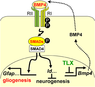
www.frontiersin.org Figure 7. A schematic diagram illustrating the interplay between TLX and the BMP-SMAD signaling pathway in the regulation of neurogenesis and astrogenesis. TLX directly modulates the expression of the BMP ligands, which bind and activate the type I (RI) and type II (RII) receptors. These events result in phosphorylation of regulatory SMADs and their dimerization with the common cofactor SMAD4. The SMAD complex acts as a transcriptional activator to induce the expression of downstream targets, which promote astrogenesis and inhibit neurogenesis.