Leta i den här bloggen


torsdag 4 juni 2015

Calreticulin ja celiakia

 Free paper abstracts The 7th International Conference on Coeliac Disease

A16
COELIAC DISEASE AUTOANTIGENS HOMOLOGOUS WITH GLIADIN
Börner H, Osman AA, Bär J, Mothes T.  University of Leipzig, Germany 
http://www.ncbi.nlm.nih.gov/pmc/articles/PMC1727378/ 

Gut. 1999 Feb; 44(2): 168–173.
PMCID: PMC1727378 Identification of common epitopes on gliadin, enterocytes, and calreticulin recognised by antigliadin antibodies of patients with coeliac disease

onsdag 3 juni 2015

Cyklofilin ja syöpä , Syklofiliini on myös molekyläärinen kaperoni.

http://www.ncbi.nlm.nih.gov/pubmed/25986562
Curr Mol Pharmacol. 2015 May 19. [Epub ahead of print]Cyclophilin function in Cancer; lessons from virus replication.

Abstract

Cyclophilins belong to a group of proteins that possess peptidyl prolyl isomerase activity and catalyse the cis-trans conversion of proline peptide bonds. Cyclophilin members play important roles in protein folding and as molecular chaperones, in addition to a well-established role as host factors required for completion of the virus life cycle. Members of the cyclophilin family are overexpressed in a range of human malignancies including hepatocellular cancer, pancreatic cancer, non-small cell lung cancer, gastric cancer, colorectal cancer and glioblastoma multiforme, however, their precise role in tumourigenesis remains unclear. In recent years, mounting evidence supports a role for prolyl isomerisation during mammalian cell division; a process with striking similarity to plasma membrane remodelling during virus replication. Here we will summarise our current understanding of the role of cyclophilins in cancer. We will review the function of cyclophilins during mammalian cell division and during HIV-1 infection, and highlight common processes involving members of the ESCRT and Rab GTPase families.
PMID:
25986562
[PubMed - as supplied by publisher]

Calreticulin, dengue , antivirusterapia ( Pohdittavaksi)

http://www.sciencedirect.com/science/article/pii/S0966842X10000624

Calretikuliiniproteiini

                     /db_xref="MIM:109091"
ORIGIN      
        1 mllsvplllg llglavaepa vyfkeqfldg dgwtsrwies khksdfgkfv lssgkfygde
       61 ekdkglqtsq darfyalsas fepfsnkgqt lvvqftvkhe qnidcgggyv klfpnsldqt
      121 dmhgdseyni mfgpdicgpg tkkvhvifny kgknvlinkd irckddefth lytlivrpdn
      181 tyevkidnsq vesgsleddw dflppkkikd pdaskpedwd erakiddptd skpedwdkpe
      241 hipdpdakkp edwdeemdge weppviqnpe ykgewkprqi dnpdykgtwi hpeidnpeys
      301 pdpsiyaydn fgvlgldlwq vksgtifdnf litndeayae efgnetwgvt kaaekqmkdk
      361 qdeeqrlkee eedkkrkeee eaedkedded kdedeedeed keedeeedvp gqakdel
//
 
 
                      /map="19p13.3-p13.2"
     Protein         1..417
                     /product="calreticulin precursor"
                     /note="Sicca syndrome antigen A (autoantigen Ro;
                     calreticulin); CRP55; ERp60; HACBP; grp60; calregulin;
                     endoplasmic reticulum resident protein 60; epididymis
                     secretory sperm binding protein Li 99n"
 signal_peptide     1..17
                     /inference="COORDINATES: ab initio prediction:SignalP:4.0"
                     /calculated_mol_wt=1693
     mature _peptide     18..417
                     /product="calreticulin"
                     /calculated_mol_wt=46467 
 
  Region          191..255
                     /region_name="4 X approximate repeats"
                     /experiment="experimental evidence, no additional details
                     recorded"
 
    Region          259..297
                     /region_name="3 X approximate repeats"
                     /experiment="experimental evidence, no additional details
                     recorded"
                     /note="propagated from UniProtKB/Swiss-Prot (P27797.1)"
 
 
 Region          414..417 (kdel) 
                     /region_name="Prevents secretion from ER" 

Miten komplementin tunnistajamolekyylin C1q reseptorit toimivat syövässä? ( cC1qR/CR and gC1qR/p33: observations in cancer)

Mol Immunol. 2014 Oct;61(2):100-9. doi: 10.1016/j.molimm.2014.06.011. Epub 2014 Jul 16. cC1qR/CR and gC1qR/p33: observations in cancer.

 Primäärituumorin elossapysyminen ja kasvu riippuu suurresti  kolmesta päätekijästä: immuunievaasiosta, angiogeneesistä ja metastasoitumisesta. Siis miten tuumori pystyy välttämään kehon immuunipuolustuksen ja miten se saa kehkeytettyä uutisverisuonia itseään varten ja miten se pääsee kehossa  etenemään.  Kehossa kasvava tuumorisolu on  "  modifioitunut oma itse"- nimittäin kehojärjestelmä tuntee oman itsensä  omilla puolustussoluillaan eikä normaalisti  tuhoa itseään- mutta tuumoreissa tämä "minä itse" on muuntunutta ja  ilmentää  suuren määrän  modifioituja pintatunnusmerkkejä ( pinta-antigeenejä) ja ne voivat  saada aikaan kehon  vasta-ainetten muodostumista, kun keho saa vihiä,  että ne eivät ole oikea  "oma itse". Nyt pitäisi seurata näistä anti-tuumorivasta-aineista  teoriassa  kehon tehokkaan komplemenettijärjestelmän aktivoituminen ja kun komplementti herää havaittuaan  "ei-itseä" olevan  solun, se käynnistää  vieraaksi tunnetun solun tuhoamisen   "ystäviensä"  kanssa ( kaikki komplementtitekijät  tekevät  tässä  sekventiaalista  yhteistyötä)   Mutta käytännössä  tämä ei  aina sujukaan.
  • The survival and growth of a primary tumor depends, by and large, on three major events: immune evasion, angiogenesis and metastasis. Tumor cells are "modified self", and as such express a plethora of modified surface antigens capable of inducing antibody production. Anti-tumor cell antibodies should, in theory, activate complement resulting in cell destruction. But this is not the case.
Syöpäsolut ovat evolutionaalisesti  ihan kuin  erilaiset patogeeniset mikro-organismit, joiden elossapysyminen riippuu isäntäsolun immuunipuolustuksen vältöstä ja ne ovat  kehittäneet  tässä evoluutiossa itselleen mekanismeja, joilla ne estävät  isäntäsoluvälitteisen solutuhon joko pidättämällä kriittisiä säätelijämolekyylejä tai kaappaamalla isäntäkehon proteiineja varmistaen  elossapysymisensä
  •  Akin to many pathogenic microorganisms whose survival depends on evading the immune system, cancer cells have also evolved diverse mechanisms to prevent host mediated cell destruction by either retaining critical regulatory molecules or by hijacking host proteins to ensure their survival.
 Kalretikuliini, joka on  tunnistavan  komplementtitekijän C1q yksi  reseptori (cC1qR)  (CR = komplementtirfeseptori)    saattaa omata  tärkää osuutta , vaikka  mainitut kolme biologista prosessia ( immuunievaasio, angiogeneesi ja  metastasoituminen)  ovatkin hyvin monimutkaisia ja  käsittävät myriadeja tuumoriin liittyviä proteiineja, entsyymejä ja sytokiinejä.
Kaikissa somaattisissa soluissa   punasolut poikkeuksena) esiintyy näitä kahta  C1q reseptorityypiä ( komplementtireseptoria)   cC1qR ( kalretikuliini)  ja gC1qR, ja ne ovat suuresti ylössäätyneitä melkein  kaikissa tuumorityypeissä. Ei ole yllättävää,    että näiden reseptorien blokeeraaminen (estäminen)  tuumorisolussa estää tuumorin kasvua;  tästä muodostuu  käsitys näiden reseptorien  tärkeydestä tuumorin kasvussa ja progredioitumsessa.
  •  Although immune evasion, angiogenesis and metastasis are complex biological processes involving a myriad of tumor associated proteins, enzymes, and cytokines, C1qRs can, nonetheless play an important role in all or part of these processes. Although both cC1qR/CR and gC1qR are expressed by all somatic cells, with the exception of red blood cells, both are highly upregulated on almost all types of tumors. It is not surprising therefore that blockade of C1qR on tumor cells inhibits their proliferation suggesting the significance of C1qRs in tumor growth and progression.
 Mielenkiintoinen havainto on myös, että näillä kahdella reseptorilla on erilaiset osatehtävät  syövän kehittymisessä (carcinogenesis):  gC1qR  edistää kasvainsolun elossapysymistä vahvistamalla angiogeneesiä ja metastasoitumista ja myös  vaikuttamalla osaltaan hyperkoagulabiliteettiä (veren  liikahyytyvyyttä) ja suosimalla mikromiljöön  tulpanmuodostusta.  Sen sijaan kalretikuliinia/ komplementtireseptoria,  cC1qR/CR ilmentävät tuumorisolut  omaavat "osoitelipun",  joka määrää ne makrofagien  tuhottaviksi.
  •  Interestingly, the two C1q receptors: cC1qR/CR and gC1qR play a differential role in carcinogenesis. While gC1qR promotes tumor cell survival by enhancing angiogenesis and metastasis and also by contributing to the hypercoagulable and prothrombotic microenvironment, cC1qR/CR expression represents a pro-phagocytic "eat-me" signal through which cC1qR/CR expressing tumor cells are tagged for destruction by macrophages.
 Tähän asti  ( lokakuu 2014)  kertyneissä tiedoissa tunnistetaan gC1qR ja cC1qR/CR  mahdollisina interventiokohteina, joihin  voidaan hahmotella  terapioita, jotka perustuvat proteiiniperusteisiin-, vasta-aineperusteisiin- tai kemoterapeutteihin, mitkä voisivat vahvistaa konventionellia syövänvastaista terapiaa.
Askeleita oikeaan suuntaan ovat todellakin  tuumorisolun proliferaation esto sellaisilla vasta-aineilla, jotka tunnistavat C1q kohdan gC1q-reseptorissa  samoin kuin  sellaisten agenssien tunnistaminen ( kuten antrasykliini), jotka tehostavat  kalretikuliini/CR ilmenemää ( cC1qR7CR expressiota)  syöpäsolussa.

  •  The data accumulated to date therefore identify gC1qR and cC1qR/CR as potential targets for the design of either protein-based, antibody-based or chemical based therapeutic intervention that could be used to enhance conventional anti-cancer therapy. The inhibition of tumor cell proliferation by monoclonal antibody recognizing the C1q site on gC1qR, as well as the identification of agents such as anthracyclin that enhance cC1qR/CR expression on tumor cells, are indeed steps in the right direction. Copyright © 2014. Published by Elsevier Ltd.

KEYWORDS: C1q receptors; C1qR and cancer; Cancer, cC1qR and gC1qR

http://www.frontiersin.org/files/Articles/23381/fimmu-03-00052-HTML/image_m/fimmu-03-00052-g001.jpg 

  Figure 1). Approximately 8% (w/w) of human C1q is carbohydrate, which is in the form of glucosylgalactosyl disaccharide units linked to hydroxylysine residues in the cC1q domain, and six asparagine-linked sugar chains located in the gC1q domain (Yonemasu et al., 1971; Calcott and Müller-Eberhard, 1972; Reid et al., 1972). The two types of sugars in turn account for approximately 69 and 31% of the total carbohydrate respectively (Mizuochi et al., 1979; Shinkai and Yonemasu, 1979; Reid, 1989).

Suomennos 3.6. 2015  

 http://www.hindawi.com/journals/bmri/2010/846872/fig1/

Kalretikuliini on lektiinin tapainen ja havaitsee sokeria.  C1q  molekyylin hiilihdyraattikohtaa, cC1q domaanin. ( Kuva)


APC funktio. Kalretikuliinin osuus MHC luokan 1 modulissa


Unlike other glycoproteins, correct folding of MHC class I molecules is not sufficient to trigger their exit from the ER, they exit only after peptide loading.

 Described here is the process of antigen presentation which consists of the folding, assembly, and peptide loading of MHC class I molecules.

 The newly synthesized MHC class I Heavy Chain (HC) is initially folded with the help of several chaperones (calnexin, BiP, ERp57) and then binds with Beta-2-microglobulin (B2M).

 This MHC:B2M heterodimer enters the peptide loading complex (PLC), a multiprotein complex that includes calreticulin, endoplasmic reticulum resident protein 57 (ERp57), transporter associated with antigen processing (TAP) and tapasin. 

 Peptides generated from Ub-proteolysis are transported into the ER through TAP. These peptides are further trimmed by ER-associated aminopeptidase (ERAP) and loaded on to MHC class I molecules. Stable MHC class I trimers with high-affinity peptide are transported from the ER to the cell surface by the Golgi apparatus.
from REACTOME source record: REACT_75795
Type: pathway
Taxonomic scope
:
organism-specific biosystem
Organism
:
Homo sapiens
BSID:
366163

Calretikuliini. Mitä uutta?


Biomed Res Int. 2015;2015:526524. Epub 2015 Mar 31.
Functional Roles of Calreticulin in Cancer Biology.
Lu YC1, Weng WC2, Lee H3. Abstract
KALRETIKULIINI on  solun hyvin konservoitunut kaperoniproteiini endoplasmisessa retikulumissa (ER) ja se osallistuu moniin soluprosesseihin.  Se tunnistettiin  ensi kerran 1974 proteiinina, joka sitoo aktiivia kalsiumjonia. 
 On alkanut kertyä näyttöä sen  suuresta impaktista   eri syöpämuotojen kehittymiseen;  sen vaikutus tuumorin muodostumisessa ja progredioitumisessa  saattaa riippua solutyypeistä ja kliinisistä vaiheista.
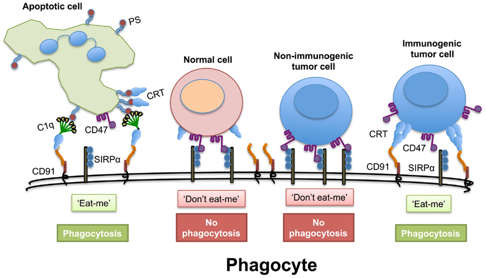
On pidetty solupinnassa sijaitsevaa kalretikuliinia solun signalointina  fagosytoiville soluille  "syö pois minut" ja niin se  edistäisi  syöpäsolujen poispoimiutumista immuunisysteemin fagosyyttien avulla.  Lisäksi on useita raportteja kalretikuliinitason manipuloimisen vaikutuksesta syöpäsolun proliferoitumiseen ja angiogeneesiin sekä solujen  erilaistumiseen.  
Immunogeenisyyden ja  tumorigeenisyyden lisäksi kalretikuliinilla on vuorovaikutuksia integriineihin solujen  adheesion aikana, mikä taas on  välttämätön prosessi kun syöpä alkaa metastasoida. 
 Integriinit ovat heterodimeerejä transmembraanisia reseptoreita, jotka yhdistävät  extrasellulaarimatrixin (ECM) ja solunsisäisen  tukirangan ( intrasellulaarisen  sytoskeletonin  solunsisäisen)    ja  liipaisevat esiin sisältä ulospäin tai ulkoa sisäänpäin johtavien signaalien kuljetuksia. 
Yhä useammat näytöt   paljastavat, että  integriiniin sitoutuvat proteiinit saattaisivat  vaikuttaa integriini-sytoskeleton- vuorovaikutusta ja sen takia  tehdä vaikutuksen solun adheesiokykyyn ( takertuvaisuuteen). 
Tässä otsikon  katsauksessa kuvataan kalretikuliinin biologisia rooleja ja tehdään yhteenvetoa kalretikuliinin mahdollisista mekanismeista, joilla se säätelee mRNA:n stabiilisuutta ja siten osaltaan  vaikuttaa syövän metastasoitumista.
  • Calreticulin is a highly conserved endoplasmic reticulum chaperone protein which participates in various cellular processes. It was first identified as a Ca2+-binding protein in 1974. Accumulated evidences indicate that calreticulin has great impacts for the development of different cancers and the effect of calreticulin on tumor formation and progression may depend on cell types and clinical stages. Cell surface calreticulin is considered as an "eat-me" signal and promotes phagocytic uptake of cancer cells by immune system. Moreover, several reports reveal that manipulation of calreticulin levels profoundly affects cancer cell proliferation and angiogenesis as well as differentiation. In addition to immunogenicity and tumorigenesis, interactions between calreticulin and integrins have been described during cell adhesion, which is an essential process for cancer metastasis. Integrins are heterodimeric transmembrane receptors which connect extracellular matrix and intracellular cytoskeleton and trigger inside-out or outside-in signaling transduction. More and more evidences reveal that proteins binding to integrins might affect integrin-cytoskeleton interaction and therefore influence ability of cell adhesion. Here, we reviewed the biological roles of calreticulin and summarized the potential mechanisms of calreticulin in regulating mRNA stability and therefore contributed to cancer metastasis.
PMID:
25918716
[PubMed - as supplied by publisher]

PMCID:
PMC4396016

Free PMC Article