Leta i den här bloggen


söndag 20 november 2016

Alkoholi ja solusykli, fork head genes

https://www.ncbi.nlm.nih.gov/pubmed/27720397
https://www.ncbi.nlm.nih.gov/pubmed/22879847 

Alcohol, fork head
 7 vastausta hakuun

Search results

Items: 7

1.
Westerhoff M, Tretiakova M, Hart J, Gwin K, Liu X, Zhou M, Yeh MM, Antic T.
Hum Pathol. 2014 May;45(5):1010-4. doi: 10.1016/j.humpath.2013.12.015.
FOXL2, a gene encoding a member of the fork-head-winged-helix family of transcription factors, is one of the earliest expressed genes during female gonadal development. It is expressed in normal ovarian stroma and ovarian neoplasms with granulosa cell lineage. Nonovarian tumors such as pancreatic mucinous cystic neoplasms (PMCs), hepatobiliary cystadenomas (HBCs), and mixed epithelial and stromal tumor of the kidney (MEST) have ovarian-type stroma. Immunohistochemical staining with FOXL2, estrogen receptor, and progesterone receptor was performed on 21 PMCs, 13 HBCs, and 10 MESTs and assessed for nuclear immunohistochemical positivity in the tumor stroma. All cases of PMC and HBC demonstrated nuclear reactivity for FOXL2 in the subepithelial stromal cells. Ninety percent of MEST demonstrated nuclear FOXL2 positivity. Estrogen receptor nuclear positivity was demonstrated in 57% of PMC, 77% of HBC, and 80% of MEST. Progesterone receptor nuclear positivity was present in 67% of PMC, 100% of HBC, and 90% of MEST. Clinical information was available for 37 patients. Seventy-eight percent of the patients had a history of obesity, heavy alcohol use, or hormone-related therapy. The 2 male patients had histories significant for morbid obesity and chronic alcoholism. FOXL2 is expressed from the early stages of ovarian development and has been shown to be mandatory for normal ovarian function. We have shown that it is also expressed in the aberrant ovarian-type stroma characteristic of PMC, HBC, and MEST. Most of such patients, including the rare male patients, have risk factors for hormonal abnormalities such as obesity and hormonal replacement therapy.
KEYWORDS:
FOXL2; Hepatobiliary cystadenoma; Mixed epithelial and stromal tumor of the kidney; Ovarian-type stroma; Pancreatic mucinous cystic neoplasm
2.
Shen L, Liu Z, Gong J, Zhang L, Wang L, Magdalou J, Chen L, Wang H.
Toxicol Appl Pharmacol. 2014 Jan 15;274(2):263-73. doi: 10.1016/j.taap.2013.11.009.

Prenatal ethanol exposure (PEE) induces dyslipidemia and hyperglycemia in fetus and adult offspring. However, whether PEE increases the susceptibility to non-alcoholic fatty liver disease (NAFLD) in offspring and its underlying mechanism remain unknown. This study aimed to demonstrate an increased susceptibility to high-fat diet (HFD)-induced NAFLD and its intrauterine programming mechanisms in female rat offspring with PEE. Rat model of intrauterine growth retardation (IUGR) was established by PEE, the female fetus and adult offspring that fed normal diet (ND) or HFD were sacrificed. The results showed that, in PEE+ND group, serum corticosterone (CORT) slightly decreased and insulin-like growth factor-1 (IGF-1) and glucose increased with partial catch-up growth; In PEE+HFD group, serum CORT decreased, while serum IGF-1, glucose and triglyceride (TG) increased, with notable catch-up growth, higher metabolic status and NAFLD formation. Enhanced liver expression of the IGF-1 pathway, gluconeogenesis, and lipid synthesis as well as reduced expression of lipid output were accompanied in PEE+HFD group. In PEE fetus, serum CORT increased while IGF-1 decreased, with low body weight, hyperglycemia, and hepatocyte ultrastructural changes. Hepatic IGF-1 expression as well as lipid output was down-regulated, while lipid synthesis significantly increased. Based on these findings, we propose a "two-programming" hypothesis for an increased susceptibility to HFD-induced NAFLD in female offspring of PEE. That is, the intrauterine programming of liver glucose and lipid metabolic function is "the first programming", and postnatal adaptive catch-up growth triggered by intrauterine programming of GC-IGF1 axis acts as "the second programming".
ACCα; ACTB; ADIPOR2; AMPKα; APOB; CORT; CPT1α; Catch-up growth; FASN; FOXO1; G6Pase; GAPDH; GC; GC–MS; GLUT2; GSK3β; Glucocorticoid–insulin-like growth factor-1 axis; HFD; HMG-CoA reductase; HMGCR; HNF4; HPRT1; IGF-1; IGF-1 receptor; IGF-1R; IGFBP3; INSR; IRS1; IRS2; IUGR; JAK2; Janus kinase 2; LEPR; MS; MTTP; NAFLD; ND; Non-alcoholic fatty liver disease; PEE; PPARα; Prenatal ethanol exposure; SREBP1c; TG; Two-programming hypothesis; acetyl-CoA carboxylase α; adenosine monophosphate activated protein kinase α; adiponectin receptor 2; apolipoprotein B; carnitine palmitoyltransferase 1α; corticosterone; fatty acid synthase; fork-head transcriptional factor O1; gas chromatography–mass spectrometry; glucocorticoid; glucose transporter 2; glucose-6-phosphatase; glyceraldehyde-phosphate dehydrogenase; glycogen synthase kinase 3β; hepatocyte nuclear factor 4; high-fat diet; hypoxanthine phosphoribosyltransferase 1; insulin receptor; insulin receptor substrate 1; insulin receptor substrate 2; insulin-like growth factor binding protein 3; insulin-like growth factor-1; intrauterine growth retardation; leptin receptor; mTORC2; mammalian target of rapamycin complex 2; metabolic syndrome; microsomal triglyceride transfer protein; non-alcoholic fatty liver disease; normal diet; peroxisome proliferator activated receptor α; prenatal ethanol exposure; sterol regulatory element binding protein-1c; triglyceride; β actin
3.
De Matteis R, Lucertini F, Guescini M, Polidori E, Zeppa S, Stocchi V, Cinti S, Cuppini R.
Nutr Metab Cardiovasc Dis. 2013 Jun;23(6):582-90. doi: 10.1016/j.numecd.2012.01.013.
 Brown adipose tissue (BAT) plays a major role in body energy expenditure counteracting obesity and obesity-associated morbidities. BAT activity is sustained by the sympathetic nervous system (SNS). Since a massive activation of the SNS was described during physical activity, we investigated the effect of endurance running training on BAT of young rats to clarify the role of exercise training on the activity and recruitment state of brown cells.
Male, 10-week-old Sprague Dawley rats were trained on a motor treadmill (approximately 60% of VO2max), 5 days/week, both for 1 and 6 weeks. The effect of endurance training was valuated using morphological and molecular approaches. Running training affected on the morphology, sympathetic tone and vascularization of BAT, independently of the duration of the stimulus. Functionally, the weak increase in the thermogenesis (no difference in UCP-1), the increased expression of PGC-1α and the membrane localization of MCT-1 suggest a new function of BAT. Visceral fat increased the expression of the FOXC2, 48 h after last training session and some clusters of UCP-1 paucilocular and multilocular adipocytes appeared.
Exercise seemed a weakly effective stimulus for BAT thermogenesis, but surprisingly, without the supposed metabolically hypoactive effects. The observed browning of the visceral fat, by a supposed white-to-brown transdifferentiation phenomena suggested that exercise could be a new physiological stimulus to counteract obesity by an adrenergic-regulated brown recruitment of adipocytes.
4.
Grønning LM, Baillie GS, Cederberg A, Lynch MJ, Houslay MD, Enerbäck S, Taskén K.
FEBS Lett. 2006 Jul 24;580(17):4126-30.
 Overexpression of forkhead transcription factor FOXC2 in white adipose tissue (WAT) leads to a lean phenotype resistant to diet-induced obesity. This is due, in part, to enhanced catecholamine-induced cAMP-PKA signaling in FOXC2 transgenic mice. Here we show that rolipram treatment of adipocytes from FOXC2 transgenic mice did not increase isoproterenol-induced cAMP accumulation to the same extent as in wild type cells. Accordingly, phosphodiesterase-4 (PDE4) activity was reduced by 75% and PDE4A5 protein expression reduced by 30-50% in FOXC2 transgenic WAT compared to wild type. Thus, reduced PDE4 activity in adipocytes from FOXC2 transgenic mice contributes to amplified beta-AR induced cAMP responses observed in these cells.
PMID:
16828089
6.
Yeon JE, Califano S, Xu J, Wands JR, De La Monte SM.
Hepatology. 2003 Sep;38(3):703-14.
Chronic ethanol consumption can cause sustained hepatocellular injury and inhibit the subsequent regenerative response. These effects of ethanol may be mediated by impaired hepatocyte survival mechanisms. The present study examines the effects of ethanol on survival signaling in the intact liver. Adult Long Evans rats were maintained on ethanol-containing or isocaloric control liquid diets for 8 weeks, after which the livers were harvested to measure mRNA levels, protein expression, and kinase or phosphatase activity related to survival or proapoptosis mechanisms. Chronic ethanol exposure resulted in increased hepatocellular labeling for activated caspase 3 and nuclear DNA damage as demonstrated using the TUNEL assay. These effects of ethanol were associated with reduced levels of tyrosyl phosphorylated (PY) IRS-1 and PI3 kinase, Akt kinase, and Erk MAPK activities and increased levels of phosphatase tensin homologue deleted on chromosome 10 (PTEN) mRNA, protein, and phosphatase activity in liver tissue. In vitro experiments demonstrated that ethanol increases PTEN expression and function in hepatocytes. However, analysis of signaling cascade pertinent to PTEN function revealed increased levels of nuclear p53 and Fas receptor mRNA but without corresponding increases in GSK-3 activity or activated BAD. Although fork-head transcription factor levels were increased in ethanol-exposed livers, virtually all of the fork-head protein detected by Western blot analysis was localized within the cytosolic fraction. In conclusion, chronic ethanol exposure impairs survival mechanisms in the liver because of inhibition of signaling through PI3 kinase and Akt and increased levels of PTEN. However, uncoupling of the signaling cascade downstream of PTEN that mediates apoptosis may account for the relatively modest degrees of ongoing cell loss observed in livers of chronic ethanol-fed rats.
PMID:
12939597

lördag 8 oktober 2016

WHO:n syöpälääkeluettelo, essentiellit.

http://www.who.int/bulletin/volumes/94/10/15-163998.pdf

Table 1.
Essential medicines for cancer on the national essential medicines lists or
national reimbursable medicines lists of 135 countries, 2015
Medicine
No. of countries listing medicine (%)
On WHO 2013 Model List
Asparaginase
68 (50)
Bleomycin
94 (70)
Calcium folinate
84 (62)
Carboplatin
73 (54)
Chlorambucil
84 (62)
Cyclophosphamide
120 (89)
Cytarabine
88 (65)
Dacarbazine
73 (54)
Dactinomycin
67 (50)
Daunorubicin
55 (41)
Docetaxel
56 (42)
Doxorubicin
99 (73)
Etoposide
84 (62)
Fluorouracil
109 (81)
Hydroxycarbamide
88 (65)
Ifosfamide
60 (44)
Mercaptopurine
87 (64)
Mesna
53 (39)
Methotrexate
128 (95)
Paclitaxel
65 (48)
Procarbazine
62 (46)
Tamoxifen
112 (83)
Tioguanine
36 (27)
Vinblastine
83 (61)
Vincristine
111 (82)
Added to WHO Model List via 2015 revision
All-trans retinoic acid
a
19 (14)
Aromatase inhibitors
b
50 (37)
Bendamustine
1 (0.7)
Bicalutamide
38 (28)
Capecitabine
49 (36)
Cisplatin
97 (72)
Fludarabine
40 (30)
Gemcitabine
46 (34)
Granulocyte colony stimulating factors
c
63 (47)
Imatinib
40 (30)
Irinotecan
41 (30)
Leuprolin class
d
53 (39)
Oxaliplatin
49 (36)
Rituximab
34 (25)
Trastuzumab
26 (19)
Vinorelbine
37 (27)
Other medicines
e
Arsenic trioxide
2 (1)
Dasatinib
10 (7)
Diethylstilboestrol
6 (4)
Erlotinib
14 (10)
Gefitinib
9 (7)
Nilotinib
12 (9

onsdag 11 maj 2016

Tiamiiniaineenvaihdunta vaikuttaa transkriptionaaliseen tasoon

http://www.ncbi.nlm.nih.gov/pubmed/23642734

J Nutr Biochem. 2013 Sep;24(9):1616-24. doi: 10.1016/j.jnutbio.2013.02.002. Epub 2013 May 1.

Up-regulation of vitamin B1 homeostasis genes in breast cancer.

Abstract

An increased carbon flux and exploitation of metabolic pathways for the rapid generation of biosynthetic precursors is a common phenotype observed in breast cancer. To support this metabolic phenotype, cancer cells adaptively regulate the expression of glycolytic enzymes and nutrient transporters. However, activity of several enzymes involved in glucose metabolism requires an adequate supply of cofactors. In particular, vitamin B1 (thiamine) is utilized as an essential cofactor for metabolic enzymes that intersect at critical junctions within the glycolytic network.

 Intracellular availability of thiamine is facilitated by the activity of thiamine transporters and thiamine pyrophosphokinase-1 (TPK-1). Therefore, the objective of this study was to establish if the cellular determinants regulating thiamine homeostasis differ between breast cancer and normal breast epithelia. Employing cDNA arrays of breast cancer and normal breast epithelial tissues, SLC19A2, SLC25A19 and TPK-1 were found to be significantly up-regulated. Similarly, up-regulation was also observed in breast cancer cell lines compared to human mammary epithelial cells. Thiamine transport assays and quantitation of intracellular thiamine and thiamine pyrophosphate established a significantly greater extent of thiamine transport and free thiamine levels in breast cancer cell lines compared to human mammary epithelial cells. Overall, these findings demonstrate an adaptive response by breast cancer cells to increase cellular availability of thiamine.
Copyright © 2013 Elsevier Inc. All rights reserved.

KEYWORDS:

Breast cancer; Metabolism; Thiamine; Transporter; Vitamin

torsdag 28 april 2016

tetraspaniini-7 proteiini PubMed hakulaiteella

COMMENT     Method: conceptual translation.
FEATURES             Location/Qualifiers
     source          1..249
                     /organism="Homo sapiens"
                     /db_xref="taxon:9606"
                     /chromosome="X"
     Protein         1..249
                     /product="tetraspanin 7, isoform CRA_a"
     Region          14..241
                     /region_name="Tetraspannin"
                     /note="Tetraspanin family; pfam00335"
                     /db_xref="CDD:249779"
     Region          110..213
                     /region_name="TM4SF2_6_like_LEL"
                     /note="Tetraspanin, extracellular domain or large
                     extracellular loop (LEL), TM4SF2_6_like subfamily.
                     Tetraspanins are trans-membrane proteins with 4
                     trans-membrane segments. Both the N- and C-termini lie on
                     the intracellular side of the membrane. This alignment...;
                     cd03161"
                     /db_xref="CDD:239414"
     Site            order(111,116,120,122..123,126,140,143..144,147..148)
                     /site_type="other"
                     /note="dimer interface [polypeptide binding]"
                     /db_xref="CDD:239414"
     CDS             1..249
                     /gene="TSPAN7"
                     /locus_tag="hCG_18324"
                     /coded_by="join(CH471141.2:1274072..1274152,
                     CH471141.2:1378638..1378826,CH471141.2:1383893..1383967,
                     CH471141.2:1386738..1386833,CH471141.2:1388222..1388377,
                     CH471141.2:1393722..1393805,CH471141.2:1400117..1400185)"
                     /note="gene_id=hCG18324.3 transcript_id=hCT1969285.1
                     protein_id=hCP1782967.1 isoform=CRA_a"
                     /db_xref="GeneID:7102"
ORIGIN      
        1 masrrmetkp vitclktlli iysfvfwitg villavgvwg kltlgtyisl iaenstnapy
       61 vligtgttiv vfglfgcfat crgspwmlkl yamflslvfl aelvagisgf vfrheikdtf
      121 lrtytdamqt yngndersra vdhvqrslsc cgvqnytnws tspyflehgi ppsccmnetd
      181 cnpqdlhnlt vaatkvnqkg cydlvtsfme tnmgiiagva fgiafsqlig mllacclsrf
      241 itanqyemv
// Essentiellit aminohapot muodostavat proteiinin alkupäässä muutamia pitkiä 
jaksoja  jopa 7 ja 6 essentielliä aminohappoa peräkkäin


Diabetes. 2016 Mar 7. pii: db151058. [Epub ahead of print]

Identification of Tetraspanin-7 as a Target of Autoantibodies in Type 1 Diabetes.

Abstract

The presence of autoantibodies to multiple islet autoantigens confers high risk for development of Type 1 diabetes. Four major autoantigens are established (insulin, glutamate decarboxylase, IA-2, and zinc transporter-8), but the molecular identity of a fifth, a 38kDa membrane glycoprotein (Glima), is unknown. Glima antibodies have been detectable only by immunoprecipitation from extracts of radiolabeled islet or neuronal cells. We sought to identify Glima to enable efficient assay of these autoantibodies. Mouse brain and lung were shown to express Glima. Membrane glycoproteins from extracts of these organs were enriched by detergent phase separation, lectin affinity chromatography and SDS-PAGE. Proteins were also immunoaffinity purified from brain extracts using autoantibodies from diabetic patients' sera before SDS-PAGE. Eluates from gel regions equivalent to 38kDa were analyzed by LC-MS/MS for protein identification. Three proteins were detected in samples from the brain and lung extracts, and in the immunoaffinity purified sample, but not the negative control. Only tetraspanin-7, a multipass transmembrane glycoprotein with neuroendocrine expression, had physical characteristics expected of Glima. Tetraspanin-7 was confirmed as an autoantigen by demonstrating binding to autoantibodies in Type 1 diabetes. We identify tetraspanin-7 as a target of autoimmunity in diabetes, allowing its exploitation for diabetes prediction and immunotherapy.
© 2016 by the American Diabetes Association. Readers may use this article as long as the work is properly cited, the use is educational and not for profit, and the work is not altered.
 

Tetraspaniiniverkosto infrastruktuuria luomassa


http://jcb.rupress.org/content/182/4/765.full

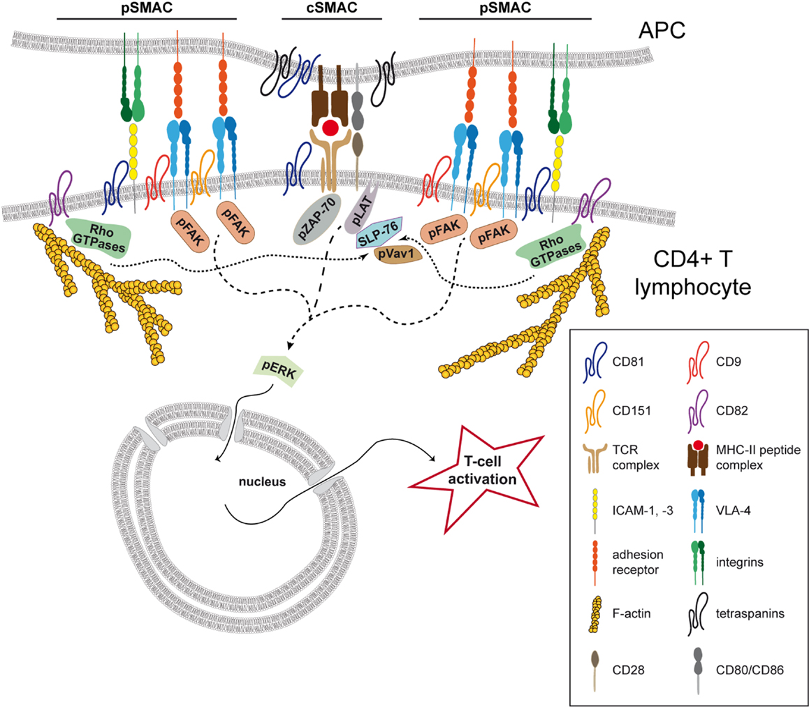
Tetraspaniinisuperperheen jäsen CD151 ja veriryhmätekijä RAPH

ISBT 025. Raph veriryhmäjärjestelmä. Geeni CD151. Kr. 11p15.5


Raph Blood Group System  Symbol RAPH; Gene locus – CD151 Alleles

Johdanto. MER2 antigeeni määrittää Raph veriryhmäjärjestelmän. (2003). Tämä antigeeni sijaitsee CD151 glykoproteiinissa, joka kuuluu tetraspaniini-proteiinien superperheeseen (TM4SP). Tetraspaniini- nimi tulee siitä, että proteiineilla on neljä konservoitua transmembraanista (TM)  jaksoa rakenteessaan. Proteiinia luonnehdittiin ensiksi trombosyyttipinnoista (GP27), mutta nykyisellään sitä on havaittu laajasti eri kudoksista ja myös punasolupinnoista. Se on adheesiomolekyyli. Kts kuva. http://atlasgeneticsoncology.org/Genes/CD151ID967ch11p15.html


      • Introduction. MER2 antigen defines the Raph blood group system. As shown recently by Crew et al. (Blood, 2003, 102, p4a) the antigen is located on CD151 glycoprotein which is a member of the four-transmembrane (TM4SF, tetraspanins) superfamily of proteins (show four conserved transmembrane domains). The protein was first characterized on platelet surface (GP27) but, has now been shown to have a wide tissue distribution and is also expressed on the surface of erythrocytes. It is an adhesion molecule.

Geenit. Geeni kromosomissa 11p15.5 on järjestäytynyt 8 exoniin ja on kooltaan 4.3 kb. Exonit 2-8 koodaavat proteiinin.

      • The genes. The gene on chromosome 11p15.5 is organized in 8 exons and spans 4.3 kb; exons 2-8 encode the protein.

Proteiinien funktio. Tämä  proteiini tekee interaktion integriineihin ja laminiiniin osallistuessaan soluadheesioon, proliferaatioon ja erilaistumiseen. Se saattaa osallistua munuaisglomeruluksen ja glomerulaarisen peruskalvon rakenteeseen ja kehitykseen. Syöpäsoluissa tämä proteiini lisää motiliteettia, invaasiota ja metastasointia.(Osuus eri  syövissä ja liittymä MMP järjestelmään ( Kirjoitan tästä enemmän MMP blogiin myöhemmin suomennosta, joka   on mainittu  linkissä  http://atlasgeneticsoncology.org/Genes/CD151ID967ch11p15.html )

      • Function of proteins. Interacts with alpha-beta integrins and laminin participating in cell adhesion, proliferation and differentiation; may be involved in the structure and development of the glomerulus and the glomerular basement membrane in man. In cancer cells enhances cell motility, invasion and metastasis.

Kudosjakauma. Proteiinia ilmenee monissa kudoksissa kuten epiteelissä, endoteelissä, lihaksessa, munuaiskeräsissä ja munuaistiehyeissä, Schwannin soluissa ja dendriittisoluissa,, fibroblasteissa ja muissa solulinjoissa.

      • Tissue distribution. Expressed in many tissues including epithelium, endothelium, muscle, renal glomeruli and tubules, Schwann and dendritic cells; fibroblasts and other cell lines.

Tautiliittymä. Eräillä yksilöillä keho ei pysty ilmentämään täyspituista CD151 proteiinia ja heillä on havaittu glomerulonefriittiä ja munuaisentoiminnan häiriötä.

      • Disease association. Failure to express full lenght CD151 was shown to be associated in some individuals with glomeluralonephritis and renal failure (Crew et al., Blood, 2004 104 2217-2223).

Alleeleista. Raph veriryhmä määräytyy yhdestä antigeenista MER2 . Sen kantajaproteiini CD 151 tunnistettiin vuonna 2003 ja sitä on useimmilla ihmisillä. Jos tämä on mutatoitunut proteiini,  yksilöt ovat MER2 negatiivisia. Heillä voi esiintyä munuaisen toiminnan vajautta  ja progressiisivtä kuuroutta, koska   myös simpukassa on samantapaisia  erikoisia "verisuoniglomeruluksia" kuin munuaisissa joiden glomerulusen peruskalvossa CD151 on tärkeä.)

  • About the alleles The Raph blood group system is defined by a single antigen MER2, first recognized by a monoclonal antibody MER2. The antigen is expressed on erythrocytes of about 92% of Caucasians but the level of expression of MER2 varies among individuals; the remaining 8% of individuals are MER2 negative.

     Crew et al. (Blood, 2003, 102, p4a) identified CD151 as the protein that carries the MER2 antigen. Sequencing of CD151 gene in four MER2 negative individuals who produced alloanti-MER2, identified specific mutations, one of which, resulting in a frameshift and truncation of the protein, occurred in three patients with renal failure. In the list of alleles the sequence acc. no. BT007397 is taken as reference.

Tetraspaniini-7

http://yle.fi/uutiset/ykkostyypin_diabeteksen_viimeinen_syy_selvisi_virittaa_toivoa_tehokkaasta_hoidosta/8840807

Brittitutkijat kertovat löytäneensä ykköstyypin diabeteksen viimeisen autoantigeenin, joka on pakoillut tutkijoita pitkään.
Diabeteksessa ihmisen oma immuunisysteemi käy hänen haimansa kimppuun ja tuhoaa sen insuliinia tuottavia soluja. Ihminen ei pysty elämään ilman veren glukoosi- eli sokeripitoisuutta säätelevää insuliinia.
Neljä immuniteettireaktion aiheuttavaa autoantigeeniä on tunnettu ennestään, viidettä on osattu kutsua vain ”glimaksi”. Nyt se on tunnistettu solukalvon proteiiniperheeseen kuuluvaksi molekyyliksi, tetraspaniini-7:ksi. Tutkimus on julkaistu Yhdysvaltain diabetesjärjestön Diabetes-lehdessä.
Tutkimusta johtanut Michael Christie kertoo Lincolnin yliopiston verkkosivulla, että gliman piilottelu tutkijoilta on haitannut diabetestestien kehittämistä. Christien tutkimusryhmään kuulunut Kerry McLaughlin jatkaa tetraspaniini-7:n tutkimusta Oxfordin yliopistossa muun muassa juuri testauksen tehostamiseksi.

http://www.lincoln.ac.uk/news/2016/04/1220.asp